A new dawn in Spatial Biology
Automating Spatial Biology with pushbutton precision
Spatial biology complexity requires next-gen precision in sample prep
Researchers struggle with labor-intensive, multistep protocols involving expensive reagents that resist scale-up and consistency. Valuable time is spent preparing precious samples rather than making life-changing discoveries.
.jpg)
Manual Sample Prep
.jpg)
scalability
%20(1).jpg)
Assay Variability
%20(1).avif)
Reagent Waste
the solution

Purpose-built Solution for Workflow Automation in Spatial Biology

.avif)
Uniform Capillary Gap Staining
Reduce reagent usage and artifacts while improving signal-to-noise

0-100 °C Precision Temperature Control
Achieve baking, HIER, and overnight incubations reproducibly

Bring Your Own Chemistries
Use our simple kits in combination with your own reagents: instrument is agnostic
.avif)
Touchscreen Protocol Builder
Create, select, edit, and customize protocols and view run reports from an intuitive interface

Plug-n-play Applications & Kits
Automate routine assays with turnkey verified protocols and single-use Skylab kits
Supported Applications
The integral solution for spatial workflows
High-content analysis methods require dewaxing, or paraffin removal, of formalin-fixed paraffin-embedded (FFPE) specimens prior to staining. High-quality dewaxing completely removes the paraffin while preserving the tissue morphology and promoting antibody binding.
Manual dewaxing is labor-intensive and inserts variability between operators and labs. Automating the process ensures complete and consistent dewaxing while you tackle burning scientific questions.
Using Parhelia Dewax reagent and kits ensures complete dewaxing without using harsh xylenes or operating under the fume hood.

The methylene bridge crosslinks introduced in formalin-fixed paraffin-embedded (FFPE) specimens must be removed so that staining probes can access target epitopes. Such epitope retrieval is achieved through heating in a buffered solution (HIER), and many HIER protocols read like cookbook recipes conducted in a kitchen rather than a scientific laboratory.
Precise heating (0-100℃) and humidity control combined with slide, coverslip, and liquid-handling automation take the guesswork out of HIER and enable hands-free protocol optimization without painstaking trial-and-error. Use the Spatial Station and Skylab kits to turn retrieval recipes into repeatable, rigorous routines.
.avif)
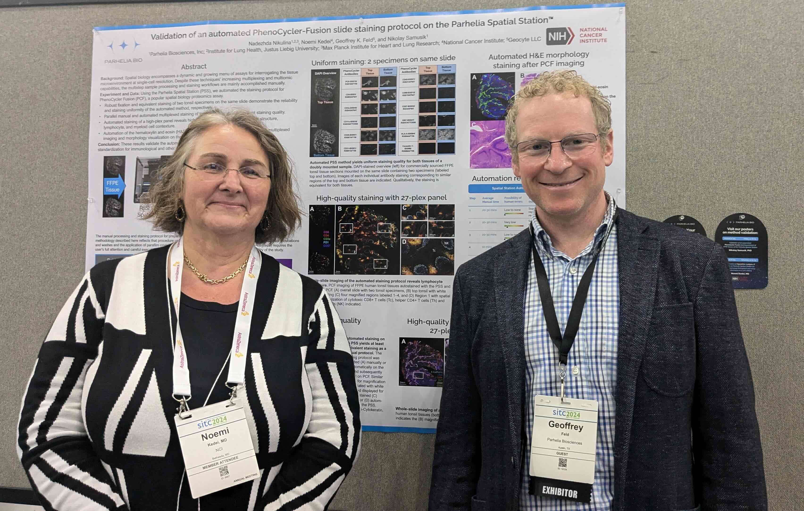
PhenoCycler technology (formally, CODEX) from Akoya Biosciences is a leading spatial proteomics assay capable of detecting 100+ targets on different tissue types at single-cell resolution. These assays are performed on PhenoCycler Fusion (PCF) systems, which automate the workflow of iterative cycles of revealing, imaging, and removing fluorescent reporters.
A series of critical staining, fixing, and washing steps must be performed before specimen imaging on PCF. The entire pre-imaging workflow has been adapted to automation on the Spatial Station with robust fixation, so you can insert your slides, push a button, and accomplish other tasks confident your samples will be stained consistently and vigorously.
For a complete end-to-end automated workflow, use the Spatial Station for:
• Dewaxing
• Epitope retrieval
• PCF staining
• Post-imaging H&E
Download the PCF White Paper
%20(2).avif)
SignalStar Multiplex IHC spatial profiling from Cell Signaling Technologies enables sensitive, accurate, and reliable data on up to 8 protein and modification biomarkers collected in just two days on FFPE samples. Researchers select and amplify IHC-validated antibody cocktails, enabling imaging of flexible, low abundance targets in tissue microenvironments.
The SignalStar workflow involves dewaxing, epitope retrieval, and precise antibody staining, amplification, and imaging incubation steps carried out at room temperature. We’ve adopted the workflow to full automation on the Spatial Station so you can design flexible biomarker studies with minimized turnaround times and manipulation of patient samples.
.avif)
GeoMx is a multiomics profiling technology whereby RNA transcripts and proteins can be spatially visualized on a GeoMx Digital Spatial Profiler (DSP) system. GeoMx is a popular tool for profiling the tumor microenvironment and uncovering predictive biomarkers. In addition to dewaxing and epitope retrieval of FFPE specimens, GeoMx requires a series of pre-imaging protocols, including proteinase treatment, fixation, probe hybridization (often occurring overnight), stringent washes, and blocking prior to H&E morphology staining.
Fortunately for multiomics spatial biologists, the Spatial Station automates the entire GeoMx pre-imaging workflow, including:
• dewaxing+HIER
•overnight staining at 4℃
•post-imaging H&E staining.
Furthermore, capillary gap methodology on the Spatial Station uses 50-70% reduced reagent with uniform staining while protecting your precious samples from delamination.
.avif)
Immunohistochemistry (IHC) is a staple assay of both research and clinical immunology labs, validating discoveries and guiding therapy choices since the 1940s. Depending on whether direct (one-step) or indirect (secondary antibody) detection is employed, sample preparation involves a series of fixation, permeabilization, blocking, incubation, and washing steps that are typically performed manually.
From dewaxing and epitope retrieval through staining, the entire IHC sample preparation procedure can be automated on the Spatial Station. Use your own buffers and antibodies and achieve reproducible, high-quality, hands-free IHC staining of any tissue sample.
.jpg)
Hematoxylin and Eosin (H&E) stain is the gold standard in medical diagnosis and pathology. Hematoxylin stains cell nuclei a purplish blue, while eosin renders the extracellular matrix and cytoplasm pink, giving a definitive contrast of the histological structure under the microscope. Often, spatial biologists will process specimens with H&E stain before or after high-content imaging, regardless of the method, adding additional manual steps to a laborious sample preparation process.
The Spatial Station quickly and reproducibly automates the H&E stain workflow, so you can combine morphology with any spatial biology analysis on any tissue without worrying about the extra steps.
Learn about our Dewax/H&E kits
.jpg)
RNAScope in situ hybridization (ISH) allows for spatial genomics, transcriptomics, and noncoding RNAs of mid-plex targets at single-molecule resolution in single cells. RNAScope builds on traditional ISH methods with additional amplification steps for improved sensitivity. While users can purchase kits and perform the assay on their own microscopes, several manual “pretreatment” blocking, incubation, and washing steps are performed at various temperatures.
With the Spatial Station, you can automate the entire pre-imaging workflow at the push of a button:
•dewaxing and epitope retrieval
•pretreatment
•probe hybridization
•amplification
• H&E staining
Customize a multiomics experiment on the same slide simply by running an additional staining protocol (e.g., PhenoCycler).
.jpg)
Immunofluorescence (IF) techniques like CycIF and Opal (from Akoya Biosciences) enable highly plexed analysis of the tumor microenvironment and single cells by combining the high sensitivity of fluorophores with the ability to combine probe cocktails sequentially. These methods are advantageous for diagnostic development since repeating the same methodology is employed for large-plex discovery as the final select panel of targets. These methods are labor intensive, as they typically require trial-and-error optimization of the dewaxing, epitope retrieval, antibody application, and removal/stripping procedures, which can limit their utility.
With the Spatial Station, you can automate the entire workflow and achieve optimal results with walkaway efficiency and consistent control over incubation times, temperatures, and staining volumes. Identify the optimum procedure automatically with slides in parallel, and then rinse and repeat to accomplish multiplexing.

Imaging Mass Cytometry (IMC) is a powerful tool for multiplexing and quantification of spatial interactions for upwards of100 protein targets. The long and tedious sample preparation workflow for IMC is manually accomplished using a hydrophobic barrier pen, which requires training, precision, and practice. Furthermore, the consecutive incubation of tissues with various buffers and reagents with different hydrophobicities that often results in leakage.
Automating the IMC sample prep protocol on the Spatial Station or Omni-Stainer makes the hydrophobic barrier pen a tool of the past. Capillary gap methodology ensures uniform and complete staining while consuming up to 5X less reagent. Most importantly, there is no risk of leakage or tissue disturbance, even during the overnight antibody incubation.
%20(1)%20(1).avif)
InSituPlex enables fast in situ visualization of up to 12 protein targets. Since the method only requires a single step for epitope retrieval, antibody staining, and subsequent amplification, InSituPlex represents a quick way to image up to 12 proteins in situ on your own microscope. Nevertheless, the method involves familiar manual processing steps including dewaxing and rehydration, HIER, blocking, staining, and incubations that require timely precision.
Users who value the speed of InSituPlex will benefit from efficient, hands-free workflow automation on the Spatial Station. Load samples, select the experimental specifics, and retrieve stained slides ready for analysis; Spatial Station handles the rest.
.avif)
Upcoming Events
Literature
Talk to our Team
Prefer email?
sales@parheliabio.com
.avif)


.avif)
